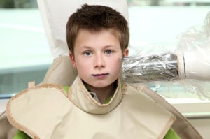
How Do X-Rays Work? Are There Different Types of X-Rays Used For My Child’s Teeth?
How dental x-rays work. X-Rays are beams of ionizing radiation. They are invisible. Ionizing radiation contains particles that carry sufficient kinetic energy to release an electron from an atom or molecule, thus converting it to an ion.
Ionizing radiation exists all around us; we absorb the radiation equivalent to 4 dental bite-wing x-rays in slightly over 14 hours just by living on Earth.
These beams are absorbed differently by various tissues in the body. They are captured on film and produce tones of gray. Each type of radiograph (x-ray) has its own advantages. Let’s look at these in detail:
Intraoral Radiographs: These are the most common. They have the advantage of providing a lot of detail. These are taken with the film inside your child’s mouth. Your child’s dentist can see tiny cavities that may be hidden, which means they can be filled before they create more harmful issues. The health of the tooth root and bone surrounding your child’s teeth also can be assessed with an Intraoral x-ray. A thorough ‘look’ at developing teeth is yet another way in which these x-rays provide helpful information.
Extraoral Radiographs: The main focus of these x-rays is the jawbone and skull. In these x-rays the film is outside your child’s mouth. Extraoral radiographs don’t provide the same detail as Intraoral radiographs, but have the advantage of showing potential issues, such as tooth impaction and improper jaw development – often while these can be more easily corrected.
Here are some examples of x-rays your child’s dentist might recommend:
INTRAORAL:
Bite-Wing X-rays will show details of the upper and lower teeth in one specific area of your child’s mouth. They reveal a clear picture of a tooth from its crown to the level of supporting bone. These x-rays can detect decay between teeth. They will also clearly show changes in bone density caused by gum disease. If your child needs a crown, a bite-wing x-ray can help her dentist measure for proper fit. The American Dental Association recommends bitewings for children only if the spaces between your child’s teeth cannot be observed during a clinical examination.
Periapical X-rays show the entire tooth – from the crown to where the tooth is anchored in the jaw, including the root. These x-rays include all teeth in one area of either the upper or lower jaw. They show the full dimension of the tooth and can be instrumental in helping your child’s dentist determine if there are abnormalities in root or surrounding bone structure.
Occlusal X-rays show full tooth development and placement, and will show the entire arch of teeth in either the upper or lower jaw.
EXTRAORAL:
Panoramic X-rays show the entire mouth – all the teeth in the upper and lower jaws – in one x-ray. This is a great tool for detecting emerging or impacted teeth, and can help with the diagnosis of tumors or other growths.
Cephalometric Projections show the entire side of your child’s head. If your child’s dentist feels it is necessary to examine his teeth in relation to his jaw, this x-ray is very useful. Often Orthodontists use this type of x-ray in order to develop a treatment plan.
Sialography allows the viewing of your child’s salivary glands by injecting a dye. This dye, called a radiopaque contrast agent, is injected directly into the salivary glands so they can be clearly seen on the x-ray film. Suspected blockages or Sjogren’s syndrome are two of the issues that would be easier to diagnose via this method.
CT Scanning. Computed tomography shows the body’s interior structures as a three-dimensional image. This is performed in a hospital, rather than a dentist’s office. Problems such as suspected tumors or fractures of the facial bones can be identified more easily by using CT Scanning.
CBCT – Cone Beam Computed Tomography uses a device that rotates around your child’s head to take many individual pictures of her jaw and teeth. These images are used to build a virtual three-dimensional representation. As a diagnostic tool, it can be the most comprehensive of all x-rays, giving a ‘whole picture’ to your child’s dentist. The advantage of a CBCT over CT Scanning is that it exposes your child to less radiation.
How dental x-rays work can best be explained by your child’s Pediatric Dentist. She has the knowledge and training to make recommendations to you about the need for x-rays. She’ll thoroughly explain the benefits and risks, and why a particular type of x-ray is best. Often dental radiographs can be instrumental in providing more accurate diagnoses; thus avoiding more invasive tests or even surgery.
Next month: Are Dental X-rays Safe for My Child? We’ll explore the pros and cons of dental x-rays and look at the facts about radiation.


